WHAT IS BREAST MRI?
Breast MRI is a non-invasive study that examines the breast tissue. Magnetic resonance imaging—MRI—uses radio signals and magnetic fields to create multiple, high-quality images.
How is a breast MRI different from a mammogram?
Mammography is the gold standard for breast cancer detection and diagnosis; however, a breast MRI can help determine the stage of breast cancer. While a breast MRI should not replace your yearly mammogram, it may be recommended by your doctor after cancer detection or if you meet certain criteria, such as a history of breast cancer or dense breast tissue.
While traditional mammography uses x-rays and compression, breast MRIs do not require compression. In contrast, using magnetic resonance imaging, these studies can detect small lesions on breast tissue that could be missed on mammograms. However, breast MRI cannot always distinguish between cancer and benign breast diseases, which can lead to false positives. Mammography remains the standard for breast cancer diagnosis and detection.
Who should have a breast MRI?
Some women might benefit from breast MRI, including those who have dense breasts. In addition, some other women might meet criteria for breast MRI, including:
- High-risk patients: The American Cancer Society guidelines recommend an annual MRI, as well as a mammogram for women who are at high risk of developing breast cancer. Heightened risk can be from a family history of breast cancer or other genetic factors, such as:
- BRCA1 or BRCA2 gene mutation
- First-degree relative with a BRCA1 or BRCA2 gene mutation
- Radiation treatment to the chest before age 30
- Li-Fraumeni, Cowden or Bannayan-Riley-Ruvalcaba syndrome (or a TP53 or PTEN gene mutation)
- ATM, CHEK2 or PALB2 gene mutation
- A greater than 20% lifetime risk of invasive breast cancer
- Breast cancer diagnosis: Women who have had cancer diagnosed in one breast should get a breast MRI to further evaluate both breasts.
- Newly diagnosed: MRI can help determine the extent and stage of breast cancer and assist in choosing the best treatment options.
- Monitoring therapy: Breast MRIs may also help monitor patients for response to treatment and to evaluate recurrent breast cancer.
- Implant status: This study is also recommended for women with breast implants to evaluate both the implant and the breast.
Breast MRI for implant integrity
While breast MRIs are commonly used for cancer detection, they can also be used for tracking breast implant integrity. The FDA has long determined silicone gel breast implants safe, though periodic imaging may be needed to ensure the integrity of implants. Currently, the FDA recommends an MRI 3 years after implantation and every 2 years to screen for implant integrity.
In addition, radiologists can determine whether an implant has ruptured with MRI imaging. Women may notice a change in breast size, hard lumps over the implant, uneven appearance, pain or tenderness, tingling, swelling, numbness or burning after a saline implant ruptures; however, women may not notice any changes when a gel implant ruptures because the material is denser. This is called a “silent rupture,” and MRI is the most effective method for detecting these ruptures.
How to prepare for a breast MRI
MRI exams are common studies that typically do not require special preparation. Here are some things to remember before your MRI:
- Bring a copy of the order for the procedure from your referring physician, a photo ID card and your insurance card.
- Take your usual medications prior to your exam.
Wear comfortable, loose clothing. Sometimes, you may be asked to change into a gown. - Before entering the MRI room you must remove ALL metallic objects including guns, hearing aids, dentures, partial plates, keys, beeper, cell phone, eyeglasses, hair pins, barrettes, jewelry, body piercing jewelry, watch, safety pins, paperclips, money clip, credit cards, bank cards, magnetic strip cards, coins, pens, pocket knife, nail clippers, tools, clothing with metal fasteners and clothing with metallic threads.
- Notify the technologist if you have any implanted electronic pacemaker, pump, stimulator or other device.
- The MRI system has a very strong magnetic field that is always on. Improper entry to the MRI scanning room may result in serious injury or death. Do not enter the MRI scanning room without the permission of the MRI technologist or Radiologist. Do not enter the MRI room if you have any question or concern regarding the safety of an implant or device.
What to expect during a breast MRI
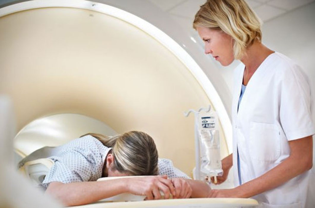
An MRI machine is a large cylindrical magnet. When you enter the MRI room, you will be asked to lie down on a bed, and the machine will move you and the bed into position, depending on the type of exam needed. During a breast MRI, you will be asked to be still for the duration of your scan, typically 20-45 minutes. Movement can blur the images and produce a lower-quality scan. The MRI machine can be loud, and you will hear loud tapping or thumping during the exam. Earplugs or earphones will be provided to you, and you can communicate with the technician through a microphone in the machine.
When your examination is over, you may resume your normal daily activities unless otherwise instructed by your doctor. One of our board-certified radiologists will review the images and send a report to your doctor.
What if I’m claustrophobic?
Some claustrophobic patients may experience a “closed in” feeling during a breast MRI. If this is a concern, you may receive a sedative, or conscious sedation, during your exam. Please let us know prior to your appointment if you think you will require a sedative. We also offer upright and open MRI options.
